Bernhard Gleich, Jürgen Weizenecker
Magnetic particle imaging (MPI) for medical diagnostics
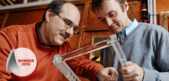
Winners of the European Inventor Award 2016
The team's magnet-based imaging method, in pre-clinical evaluation since 2014, promises to enable doctors to obtain instant, three-dimensional images of soft tissue complications, including cancers and vascular diseases, with a spatial resolution of up to 0.5 millimetres. MPI also unlocks applications in materials sciences and fluid dynamics, as well as new levels of quality and safety control by detecting surface cracks and fractures.
Bernhard Gleich and Jürgen Weizenecker achieved their breakthrough by extensively studying the properties of superparamagnetic iron oxide nanoparticles (SPIONs) in response to an oscillating magnetic field. The interaction between magnetic field and particles, administered to patients in liquid form and safely processed via the body's iron metabolism, offers the potential to mapping arterial pathways and organs in 3D during a wide range of dynamic medical phenomena.
Societal benefit
Coronary heart disease (CHD) is a major cause of death and disability in developed countries, currently accounting for over one third of deaths in people over 35 years of age (American Heart Association). Prevention and early diagnosis are important for stopping the often slowly progressive disease that will affect nearly one half of all middle-aged men and one third of middle-aged women at some point.
The clinical availability of MPI would indicate a new level of mapping coronary pathways and detecting arterial blockages. In addition to detecting cancers, MPI could also deliver live images during surgery, affording physicians insights into the effects of manipulation or drug injections in real time.
Economic benefit
MPI technology entered into pre-clinical trials in September 2014, when Philips' partner company, Bruker Corporation, installed its first pre-clinical MPI scanner at the University Medical Center Hamburg-Eppendorf (UKE), Germany. Among other applications, researchers use the scanner for angiographic tomography, drawing on its excellent capacities to image the cardiovascular system. Headquartered in Billerica, Massachusetts, USA, with 6 100 employees and EUR 1.65 billion in revenue (2014), Bruker Corporation specialises in scientific instruments for molecular and materials research.
The fast and precise scanning devices could cause a shift in the segment of the Pre-clinical Imaging (in-vivo) market, currently forecast to reach EUR 731 million globally by 2019, at a compound annual growth rate (CAGR) of 6.0% from 2014 to 2019 (MarketsandMarkets).
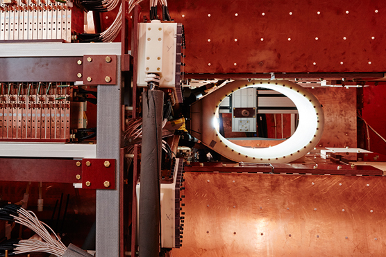
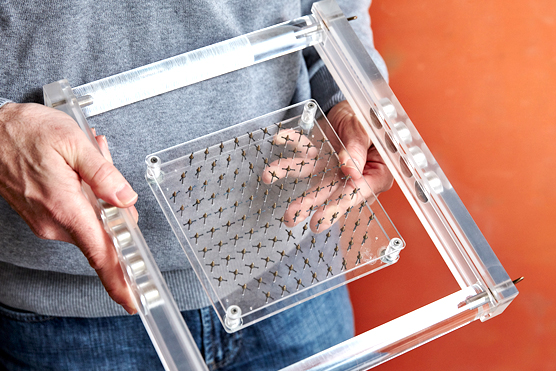
How it works
MPI relies on a two-step principle: Before examination, patients ingest a nano liquid with miniscule superparamagnetic iron oxide nanoparticles (SPIONs) measuring between 20 and 40 nanometres. In the second step, these magnetic particles are made detectable by applying a magnetic field - known as a drive field - which gives them a magnetic resonance of their own, rendered by 3D-imaging software as spatial maps of the body.
Through their magnetic charge, SPIONs deliver detailed images of soft tissue and vascular pathways that are difficult to obtain with other methods such as MRI, which measures water in the human body. The term "superparamagnetic" means that SPIONs hold no further magnetisation once the magnetic field is deactivated, after which they are safely processed by the body's metabolism.
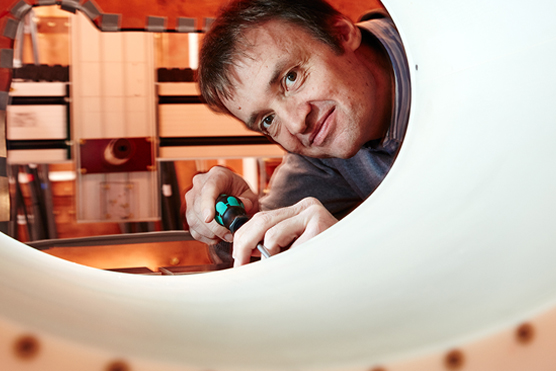
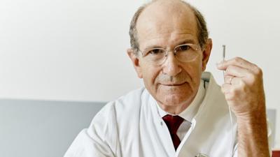
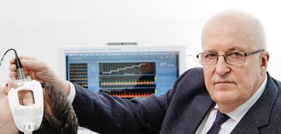
The inventors
Bernhard Gleich and Jürgen Weizenecker both worked independently on the principles of magnets and paramagnets - Gleich at the University of Ulm and Weizenecker at the University of Karlsruhe - before joining Philips Research Hamburg in 2000. In 2005, the two German physicists published the method behind MPI in Nature magazine as a promising new method for detecting vascular diseases and cancers.
Over the years, the two inventors have been granted 18 joint patent families from the European Patent Office for improving MPI, including a superposed oscillating magnetic field yielding signals in even greater detail.
Gleich's thesis entitled "Principles and Applications of Magnetic Particle Imaging" earned him a PhD from the University of Lübeck in 2013 and the coveted Professor-Otto-Rohe Award in 2014. He is currently pursuing work on an MPI prototype scanner. Weizenecker, who has been a professor of Electrical Engineering and Information Technology at the University of Applied Sciences in Karlsruhe since 2008, is pursuing translational research in MPI.
Did you know?
At first sight, the iron oxide nanoparticles used in MPI may not look like the kind of agent any patient would like to drink or receive via injection. But iron oxide particles are basically just that - iron. This makes them perfect candidates for being broken down via the body's iron metabolism within a matter of hours or days.
In fact, iron is one of the keys to life: All of the body's cells require iron to function, as iron is a crucial element in energy production, oxygen transport, and cellular growth. The human body contains around 3.5 grams of iron on average, and the typical diet adds a daily intake of 10-20 mg. Only 10% of dietary iron is absorbed; the excess is transported by a glycoprotein called transferrin. Transferrin is used to handling far larger workloads than MPI's miniscule amounts of iron oxide.
Media gallery

- Magnetic Particle Imaging (MPI)\".")
\".")
Related finalists


Contact
European Inventor Award and Young Inventors Prize queries:
european-inventor@epo.org Subscribe to the European Inventor Award newsletterMedia-related queries:
Contact our Press team#InventorAward #YoungInventors