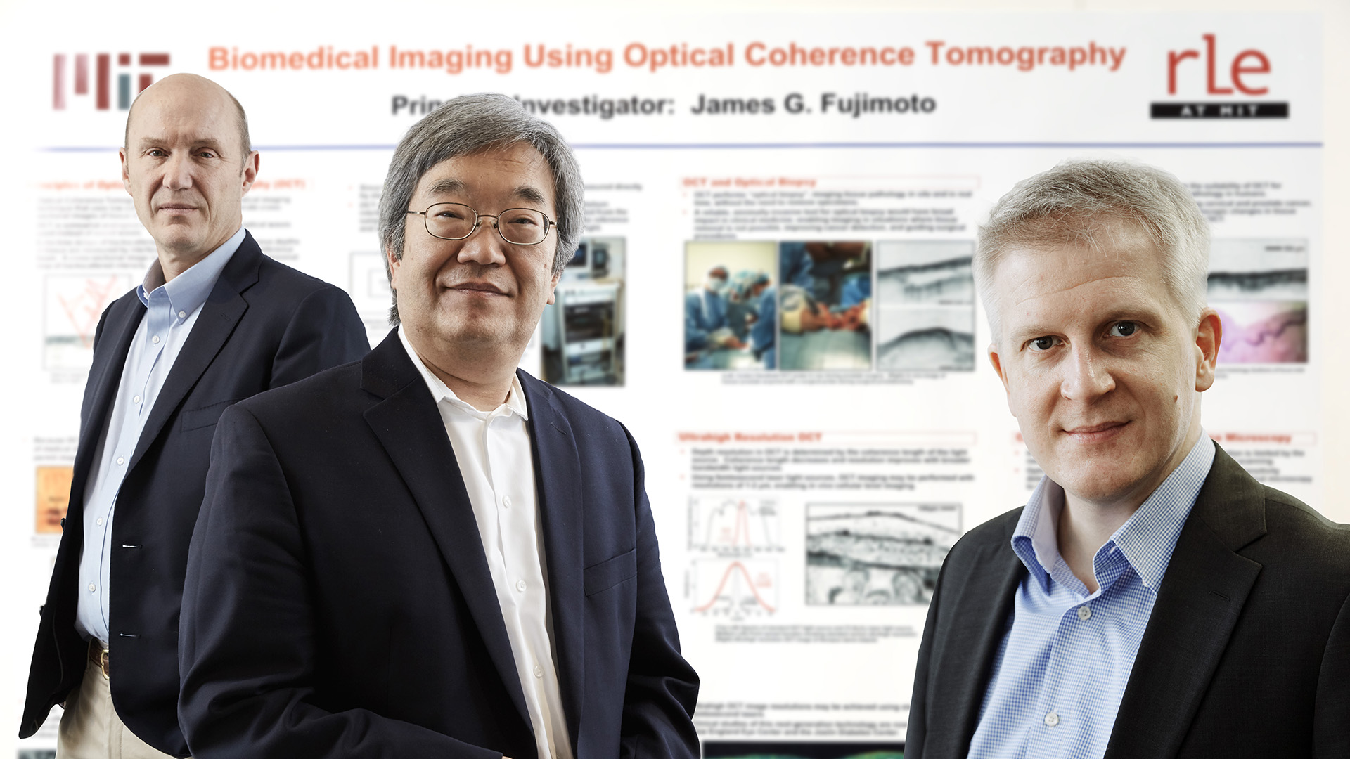
James G. Fujimoto, Eric A. Swanson and Robert Huber
Medical imaging with optical coherence tomography (OCT)
 (ceremony)\".")
Winners of the European Inventor Award 2017
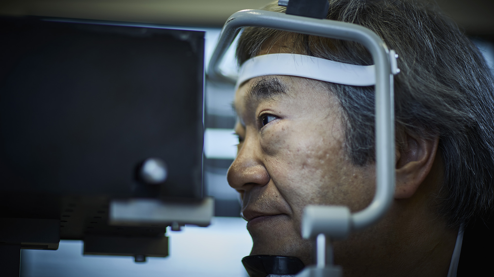
Premiered as a clinical prototype in 1993, OCT solves a long-standing problem in diagnostic medicine: accurate evaluation of soft body tissue and blood vessels - key to diagnosing cancer and cardiovascular diseases - without invading the body. Biopsies have long been used, but they require tissue samples from the area in question and have never been considered an ideal method. The first imaging method of its kind, OCT creates an optical biopsy: real-time, 3-D images of soft tissue at microscopic resolutions.
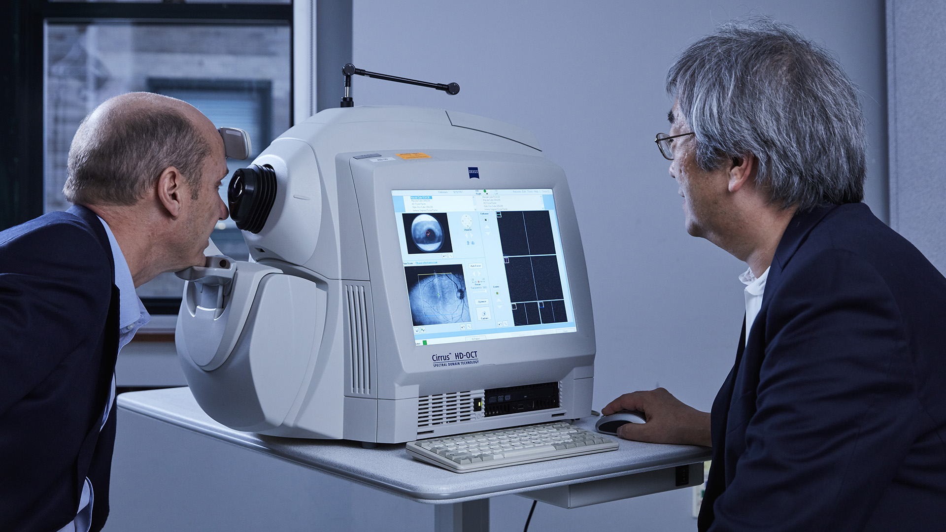
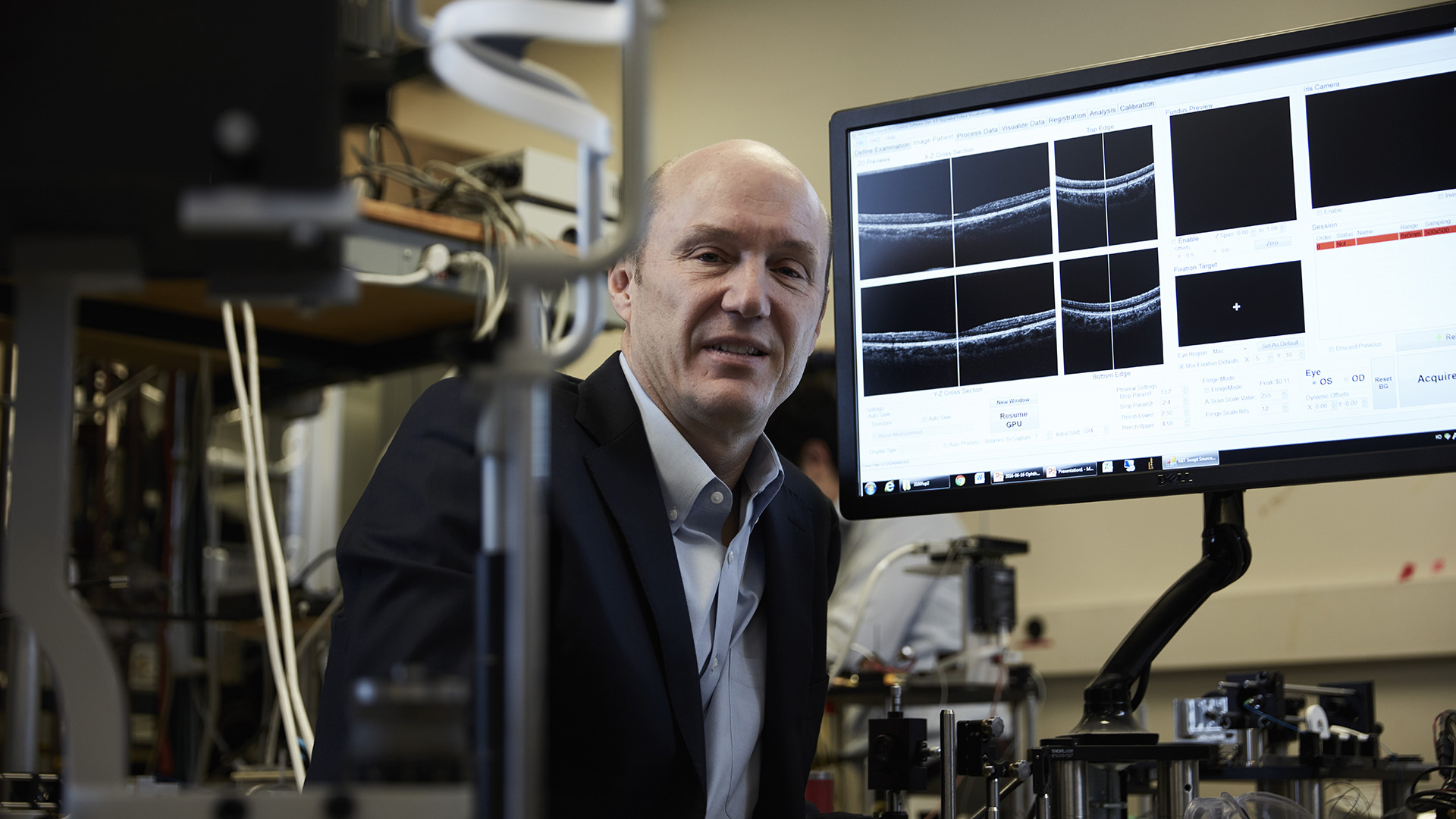
Fujimoto and Swanson started developing the technology in 1990 at the Massachusetts Institute of Technology (MIT) in Boston, ultimately filing more than 50 patents in the process. Their breakthrough was to aim a laser at soft body tissue and measure the "echo", i.e. the time delay of the light beams. Originally developed for ophthalmology, OCT has become the standard for eye care, with 30 to 40 million imaging procedures performed each year.
After commercial OCT devices for ophthalmology became available in 1996, the technology was adapted to other medical fields: cardiovascular OCT scanners appeared in 2004, followed by dermatological OCT in 2010 and gastrointestinal OCT in 2013.
Societal benefit
Glaucoma is the second leading cause of blindness worldwide, with current studies estimating the number of suspected cases at over 60 million. Early diagnosis - now possible with OCT - is the key to preventing further loss of vision. The technology has given doctors a new tool to diagnose serious eye diseases at early, treatable stages, preventing vision loss and serious complications in countless cases.
Through its widespread availability and coverage by major insurance companies, OCT has helped reduce costs for patients and caregivers. In newer applications such as gastroenterology, virtual endoscopy of the gastrointestinal tract using OCT could save over EUR 600 million per year. The technology's capacity for live imaging during surgery has increased safety in the operating room. It has contributed to pharmaceutical drug development and advanced understanding of the cause of diseases.
OCT also unlocks new levels of precision in genetic research, materials quality control, art conservation and optical data storage.
Economic benefit
Key technologies developed and patented by Fujimoto and his team were acquired by optical and optoelectronics firm Carl Zeiss, currently a market leader in the field. In fiscal 2015/2016, the company's Ophthalmic Systems unit - home to OCT solutions - generated revenues of EUR 421 million, a 7.5% increase over the EUR 392 million revenue of the previous year.
The team founded start-ups of its own, including Advanced Ophthalmic Devices in 1992 and LightLab Imaging in 1998 (Fujimoto and Swanson), and Optores GmbH in 2013 (Robert Huber). The invention has spawned a thriving market with considerable start-up activity: the OCT industry employs roughly 16 000 professionals, ranging from engineers to hospital staff. Over 100 companies currently supply OCT systems and components, and more than 50 000 clinical OCT systems have been installed worldwide.
A true blockbuster technology, OCT created total revenues upwards of EUR 4.77 billion between 1996 and 2016. Analysts at BioOpticsWorld estimated global revenues from OCT systems in 2015 at EUR 688 million. Experts at Research and Markets expect the OCT market to grow at a compound annual growth rate of 12%, ultimately reaching over EUR 1.5 billion by 2020.
How it works
Before the arrival of OCT, scientists lacked adequate methods to diagnose abnormalities in soft tissue. These included tumours of the eye and stomach, as well as obstructions in blood vessels not visible with X-ray or magnetic resonance imaging (MRI).
The principle behind OCT is similar to ultrasound. But instead of sound waves, the system uses the "echo" of light beams refracted from human tissue. The technology requires no contrast fluids or preparation of body tissue.
An OCT procedure takes a few minutes and works in three steps. A fast-pulse laser shoots near-infrared light into soft tissue about 1-2 millimetres deep. The scattered light from the tissue reveals anatomical features when compared to a reference beam of unscattered light. Finally, data from the light beams is processed by computer software and rendered into images with microscopic clarity.
The inventor
Fujimoto earned his PhD in Electrical Engineering and Computer Science from MIT in 1984 and still teaches at his alma mater. Currently a principal investigator at MIT's Research Laboratory of Electronics (RLE) and Department of Electrical Engineering and Computer Science, Fujimoto is listed as inventor or co-inventor on 15 patent families. He has contributed to over 450 journal articles and nine books. For his contributions to medical imaging and ophthalmology, Fujimoto has been honoured with numerous international awards, such as the Carl Zeiss Research Award (2011), the António Champalimaud Vision Award (2012), the IEEE Photonics Award (2014), the Optical Society's Frederic Ives Medal (2015) and the National Academy of Engineering's Russ Prize (2017).
Eric Swanson earned his Masters of Science in electrical engineering from MIT in 1984. Co-author of 81 journal articles, 142 conference presentations and over 40 patents, he has co-founded several start-ups, including Coherent Diagnostic Technologies (later LightLab Imaging) in 1998 with Fujimoto as the world's first OCT supplier for cardiology applications. Swanson's contributions have been honoured with numerous awards, including the Rank Prize in optoelectronics (2002), the António Champalimaud Vision Award (2012) and the National Academy of Engineering's Russ Prize (2017).
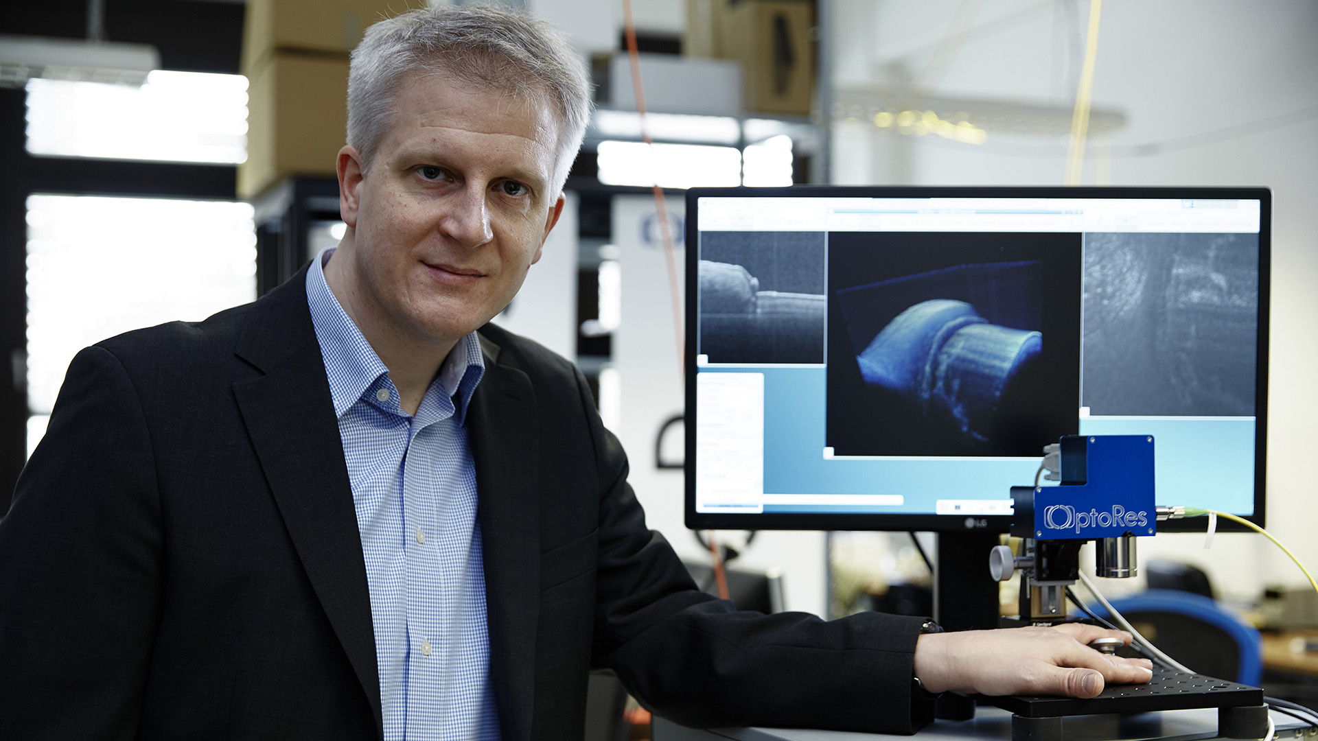
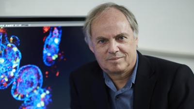
Laser specialist Robert Huber earned his master's degree at the Faculty of Physics of Munich's Ludwig-Maximilians-Universität (LMU) in 1998 and his PhD in 2002. He is the author of 100 peer-reviewed publications, is listed as inventor on seven European patent applications and has received two ERC grants from the European Union. Currently a professor at the Institute for Biomedical Optics at the University of Lübeck, Germany, Huber co-founded the Munich-based company Optores GmbH in 2013 to develop an ultra-fast version of OCT. Huber has been honoured with the German Chemical Society's Albert Weller Award (2003), the Rudolf Kaiser Award (2008) and the Klung Wilhelmy Weberbank Award (2013).
Did you know?
Focused light from devices as small as a hand-held laser pointer can have devastating effects on the eye. In 2015, the US Food and Drug Administration (FDA) reported over 5 000 eye injuries involving laser pointers. FDA regulations limit the visible light power of hand-held laser pointers to 5 milliwatts.
The dangerous potential of lasers did not deter Fujimoto from volunteering as the first human test subject for the OCT prototype apparatus in 1993. He showed up, removed his glasses and declared himself ready to have the apparatus shine a laser light directly into his eye. The OCT device proved to be safe and has since advanced to become the world's most commonly used imaging technology in ophthalmology.
Media gallery
\".")
Related finalists
Contact
European Inventor Award and Young Inventors Prize queries:
european-inventor@epo.org Subscribe to the European Inventor Award newsletterMedia-related queries:
Contact our Press team#InventorAward #YoungInventors